Research Highlights
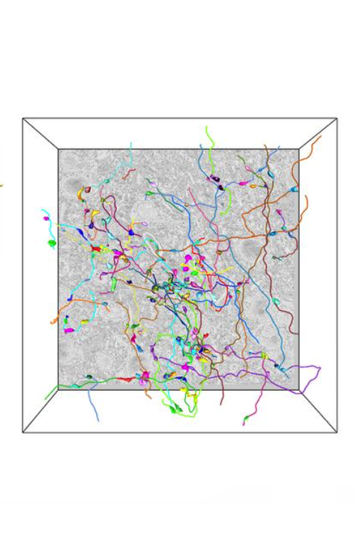 |
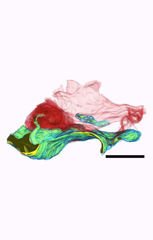
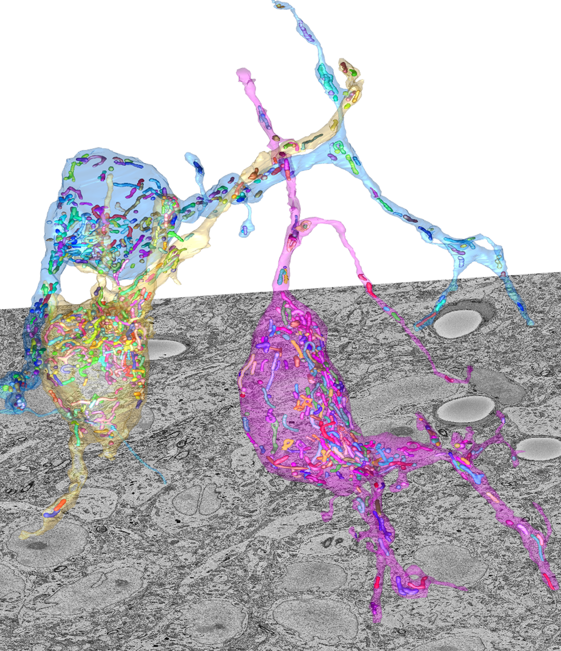
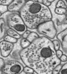
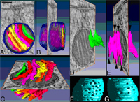
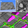
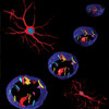
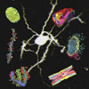
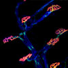
Ultrastructure of Synaptic Connectivity within Subregions of the Suprachiasmatic Nucleus Revealed by a Genetically Encoded Tag and Serial Blockface Electron MicroscopyInvestigators form the Salk Institute for Biological Studies partnered with NCMIR to apply advanced EM techniques to study the connectivity of hypothalamic suprachiasmatic nucleus (SCN) neurons. The research reveals differences in synaptic input, dendritic network density, and criteria for identifying various axonic boutons within the SCN. Read More > > |
 |
Photoreceptor Disc Incisures Form as an Adaptive Mechanism Ensuring the Completion of Disc EnclosureInvestigators from Duke University leverage NCMIR’s expertise in multi-tilt STEM tomography to help characterize the ultrastructure and development of key constituents of retinal photoreceptor cells. Read More > > |
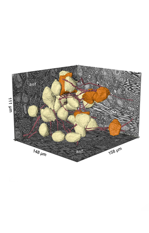 |
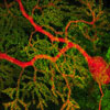
High-resolution volumetric imaging constrains compartmental models to explore synaptic integration and temporal processing by cochlear nucleus globular bushy cellsProfessor George Spirou from the University of South Florida led an interdisciplinary consortium of investigators in the structural analysis of bushy cells in the mouse cochlear nucleus. The team, which included researchers from NCMIR, the University of North Carolina, and West Virginia University, combined advanced volume electron microscopy techniques with sophisticated computational modelling to predict heterogeneous bushy cell responses. Read More > > |
 |
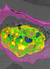
Scientists Discover Key Information About the Function of Mitochondria in Cancer CellsScientists have long known that mitochondria, the "powerhouses" of cells, play a crucial role in the metabolism and energy production of cancer cells. However, until now, little was known about the relationship between the structural organization of mitochondrial networks and their functional bioenergetic activity at the level of whole tumors. Read More > > |
 |
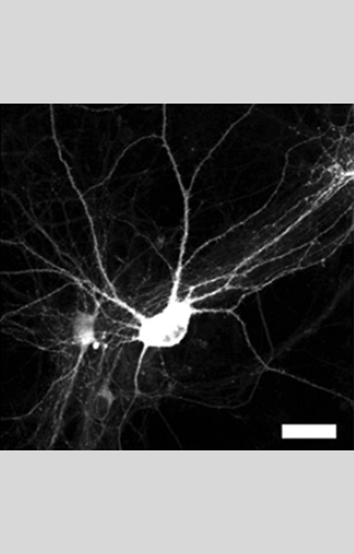
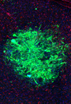
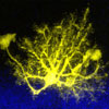
A New Framework for the Formation and Development of Learning-related Dendritic SpinesUCSD Investigators in the laboratory of Professor Takaki Komiyama partner with NCMIR to deepen understanding of the basics of information processing in the brain, and how such processing can flexibly change with learning. Correlated light an electron microscopy was used to study the generation and development of learning-related dendritic spines, revealing new insight into the degree to which activity, location, and potentiation of existing spines affected the growth and maintenance of new spines. Read More > > |
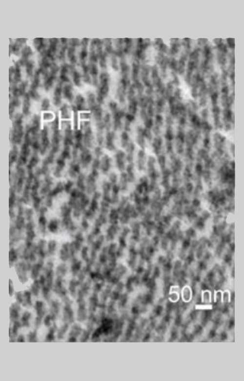 |
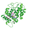
Distinct tau neuropathology and cellular profiles of an APOE3 Christchurch homozygote protected against autosomal dominant Alzheimer's dementiaNCMIR researchers join a consortium of investigators studying an autosomal dominant Alzheimer’s disease (ADAD) PSEN1 E280A carrier who was also homozygous for the APOE3 Christchurch (APOE3ch) variant and was protected against Alzheimer’s symptoms for almost three decades beyond the expected age of onset. Work reported in Acta Neuropathologica offers new insight into the Christchurch variant’s role in the distribution of tau pathology and the severity, progression, and clinical presentation of Alzheimer’s Disease, suggesting possible therapeutic strategies. Read More > > |
 |
Long-lived Proteins in Mitochondria of the Brain Stabilize Protein ComplexesMitochondria are known as the powerhouses of the cell, generating the energy that’s needed to fuel the functions that our cells carry out. Now, scientists at the Salk Institute and the National Center for Microscopy and Imaging Research (NCMIR) have taken a closer look at how mitochondria are maintained in nondividing cells, such as neurons, with the ultimate goal of developing a better understanding of how to prevent or treat age-related diseases. The researchers found that many of the proteins in mitochondria last much longer than expected, and that this stability likely protects them from damage. The findings were published October 28, 2021, in Developmental Cell. Read More > > |
 |
Overcoming Obstacles to Promote Repair in Multiple SclerosisFindings from a collaborative study conducted by scientists at the Gladstone Institutes, led by Senior Investigator Katerina Akassoglou, PhD, NCMIR and the University of Vienna, were recently published in the journal Brain. This team explored the efficacy of drugs, currently in trials, for promoting the repair of myelin damage associated with multiple sclerosis. They identify a new line of therapeutics with the potential to stimulate myelin repair even in the presence of toxic blood leaks in the brain. Read More > > |
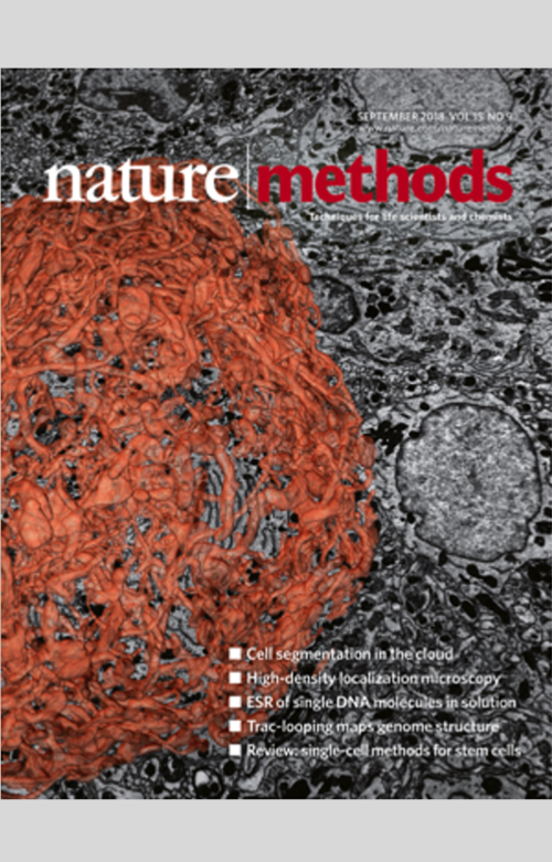 |
CDeep3M—Plug-and-Play cloud-based deep learning for image segmentation
In the September 2018 issue of Nature Methods, NCMIR researchers debut a ready-to-use image segmentation solution for any lab. The system, called CDeep3M, includes a pre-configured and publicly available deep convolutional neural network that can be run on the Amazon cloud compute platform. Read More > > |
 |
High-performance serial block-face SEM of non-conductive biological samples enabled by focal gas injection-based charge compensationIn collaboration with Carl Zeiss Microscopy (www.zeiss.com), NCMIR researchers developed a new technology for improving the performance of serial blockface scanning EM (SBEM) using the Gatan 3View. This technology allows for the focused and synchronized delivery of nitrogen gas to the sample blockface during the imaging cycle to eliminate charging, greatly extending the utility of the volume imaging technique for even the most-charge-prone samples. Read More > > |
 |
Intrinsic Assembly of Neural Circuits
Scientists at The Scripps Research Institute (TSRI) and the National Center for Microscopy and Imaging Research (NCMIR) in La Jolla have revealed new clues to the wiring of the brain. A team led by Associate Professor Anton Maximov, in collaboration with Dr. Mark Ellisman’s group, have found that neurons in brain regions that store memory can form networks in the absence of synaptic activity. Read More > > |
 |
Brain Synapse Size Grows Larger, Then Smaller in Regular Cycle Between Wakefulness and SleepA team of researchers at the University of Wisconsin-Madison and UCSD/NCMIR discovers that synapses in mouse cerebral cortex strengthen (grow larger) during wake to support learning but decline in strength (grow smaller) during sleep to consolidate memories and restore cellular function. Read More > > |
 |
Multicolor Electron Microscopy Used to Visualize Multiple Molecular Components Inside a CellThis method applied different lanthanides to label specific cellular components. The lanthanides’ electron energy-loss spectra were visualized, and the resulting elemental maps overlaid on a conventional greyscale EM image to “color-code” the labeled components and enable tracking of their movements over time across multiple images. Read More > > |
 |
NCMIR Develops New Technique to Improve Resolution of SBEM ImagesApplying a negative voltage to an SBEM stage creates a reversal potential that decreases the landing energy of probe-beam electrons. At a lower landing energy, probe electrons penetrate more shallowly, resulting in improved resolution. The reversal potential further accelerates backscattered electrons, leading to a large increase in their recorded signal by the detector. Read More > > |
 |
Using Click-EM for Metabolic Labeling to Track and Image Tagged Non-protein Biomolecules by Light and Electron Microscopy
UCSD and NCMIR researchers introduce a powerful new approach for metabolic labeling and imaging of non-proteinaceous cellular components by light and electron microscopy. Read More > > |
 |
Advances in Molecular Labeling Tools Enhance Results Using Multiscale, Multimodal, Correlated Microscopies
Scientists at the National Center for Microscopy and Imaging Research (NCMIR) have summarized four recently developed, now widely used, electron microscopy (EM) probes (also called labels or tags) in a review to facilitate more general adoption of new probes and methods from the center. Read More > > |
 |
CNS Myelin Turnover Mechanism Clarified: Astrocytes Eliminate Cellular Debris Resulting from Removal of Myelin Segments as the Optic Nerve Shortens During Frog Metamorphosis
Researchers at The Johns Hopkins University School of Medicine, the Hugo W. Moser Research Institute at Kennedy Krieger, Inc., and the National Center for Microscopy and Imaging Research demonstrated that, in a developing vertebrate ON, individual myelin segments can shorten in response to shortening in axon length, revealing a plasticity of myelin segments not previously known. Through volume EM-based quantification, the team further demonstrated that axon myelin segments shorten through selective astrocytic phagocytosis of small intra-segmental myelin dystrophies. Read More > > |
 |
Remodeling of the Nuclear Pore Complex by the p75 Neurotrophin Receptor Controls Transforming Growth Factor Signaling and Astrocyte Functions
Researchers from UC San Francisco, the University of Freiburg (Germany), UC San Diego and its National Center for Microscopy and Imaging Research, the University of Glasgow (UK), King’s College London, and Stanford University team to establish a potential, previously unknown, mechanism by which the brain responds to injury. Read More > > |
 |
New Insight Toward Understanding the Molecular Basis for Long-term Memory
Human memory can persist for a lifetime, yet the majority of synaptic proteins in the brain turn over at a rate of hours to days. Synthesizing, localizing, and stabilizing new protein copies at synapses are crucial factors in maintaining the synaptic changes required for storing long-term memories. To gain insight into how long-lasting changes in synaptic strength are sustained, a research team from UC San Diego and the Howard Hughes Medical Institute investigated PKMζ and PKCλ stability in rat neurons. Read More > > |
 |
New Experimental Protocols for Correlative Stochastic Optical Reconstruction Microscopy and EM
A research team from the Howard Hughes Medical Institute, Harvard University, the National Center for Microscopy and Imaging Research, and UC San Diego developed a new method that combines 3D stochastic optical reconstruction microscopy (STORM) with several EM imaging modes, including protocols to image unembedded and embedded/sectioned samples. They applied the resulting protocols to several cellular and viral structures. Read More > > |
 |
APEX2 Enhances Proximity Labeling and Electron Microscopy Imaging to Resolve Dispute about Regulation of the Inner Membrane ChannelA research team from MIT, Massachusetts General Hospital, and UCSD in late November 2014 published a study in Nature Methods describing an electron microscopy tag, APEX2, which they developed for live-cell proteomics and genetic probe-based labeling for light and electron microscopy. Use of this tag enabled them to resolve a conflict between competing mechanistic models of regulation of the inner membrane channel through which calcium is supplied to mitochondria. Read More > > |
 |
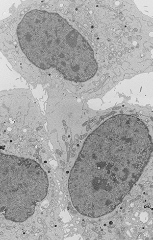
Automatic Segmentation of Organelles in Electron Microscopy Image Stacks: A New WorkflowElectron microscopy is a very useful technique to analyze the form, distribution, and functional status of key organelle systems in various pathological processes including those associated with neurodegenerative diseases. Significantly, it has been used to provide important new insights into the mechanisms underlying diseases such as Parkinson’s and Alzheimer’s diseases and glaucoma.Moreover, recent advances in EM instrumentation are fueling a renaissance in the study of quantitative neuroanatomy. Data obtained from techniques such as serial block-face scanning electron microscopy (SBEM) provide unprecedented volumetric snapshots of the in-situ biological organization of the mammalian brain across a multitude of scales. Read More > > |
 |
X-ray Microscopy Increases Accuracy and Efficiency of SBEM Imaging for Correlated Light/Electron Microscopy of Biological SpecimensThe recently developed 3D method of serial block-face scanning electron microscopy is rapidly establishing itself as a powerful imaging approach in the biological sciences research community. This approach is proving of great value for 3D visualization of nervous system ultrastructure, particularly when details of synaptic and other subcellular elements must be located and quantified. It is also important for constructing connectomic models of regional brain circuits. In addition, SBEM is increasingly used to perform nanohistology on a wide variety of tissue systems, including the lung, liver, tendons, kidney, and cell cultures. Read More > > |
 |
Epik! A New Scientific Workflow for Electron TomographyComputational researchers, regardless of their disciplines, need software tools that save time, optimize and scale up computations, produce results faster, create an extensible platform, foster collaborations, effectively communicate the underlying science, and enable others to replicate the results. Some of the most effective tools are based on scientific workflows. A workflow is a software application that solves a scientific problem. It’s composed of computational steps and data manipulation tools that can scale up to run on high-performance computers, distributed environments, and commercial cloud systems. Workflows are used in all stages of the data lifecycle: generation and acquisition, analysis, comparison, publication, and archiving. Read More > > |
 |
PRIME Directive: New Chemical Fluorophore Designed to Tag Cell Proteins for ImagingFluorescent proteins are used commonly to tag components of interest in living cells for subsequent imaging. But there’s a cost: Their dim fluorescence, rapid photobleaching, and large size limit their utility, and they can disrupt protein folding and trafficking and/or impair protein function.Chemical fluorophores, by comparison, tend to be considerably smaller, brighter, and more photostable, characteristics that allow them to perform better in advanced biological imaging modalities such as single-molecule tracking and super-resolution microscopies. However, it is much more challenging to target them to specific cellular proteins inside living cells because they cannot be genetically encoded and have to target proteins post-translationally inside a complex cellular environment. Read More > > |
 |
Astrocytes Devour Neuronal Mitochondria in a Newly Documented Process Called Transmitophagy
Scientific canon long held that when the tiny energy producers within cells, called mitochondria, become damaged or dysfunctional, these tiny organelles are degraded and recycled within the cell that produced them. But a paper appearing in the Proceedings of the National Academy of Sciences (PNAS) from The Johns Hopkins University and NCMIR scientists now shows that, instead, particularly in the long axons of the brain’s nerve cells, damaged mitochondria are transferred to nearby glial cells for elimination. This process, in effect, outsources a substantial portion of the neurons’ housekeeping. Read More > >
|
 |
A New Model System for Neural Development Produces Nanoscale-resolution Animations that Inspires Music
Neural circuit development in mice is characterized by early exuberant innervation followed by competition and pruning to mature innervation topography. Several neural systems (e.g., the neuromuscular junction ad climbing fiber innervation of Purkinje cells) have served as models to study neural development in part because they establish a recognizable endpoint of mono-innervation of their targets, and the presynaptic terminals are large and easily monitored. The calyx of Held (CH) innervation of its target, which forms a key element of auditory brainstem binaural circuitry, exhibits these same characteristic. Read More > >
|
 |
De-coating Caveolae: Understanding the Caveolar Protein Machinery in Endocytosis
Caveolae are flask-shaped invaginations with a diameter of 50-80nm in the plasma membrane of many types of mammalian cells. They are particularly abundant in fat cells, muscle cells, and cells that line blood vessels and serve as a source for clathrin-dependent endocytosis. Although they are likely to be important for cellular responses to mechanical stress, intracellular trafficking, and signaling events, the precise molecular mechanisms that determine how they form and carry out these functions have yet to be understood. Read More > >
|
 |
Three Types of Microscopy Go To the Limit to Determine Ryanodine Receptor Calcium Release Channels in Cardiac Myocytes
A research team led by UCSD recently published results of their work using various kinds of microscopy to reveal the relative distribution of ryanodine receptors and caveolin-3 in mouse ventricular myocytes in the cytosol and near the cell surface. Ryandine Receptors are important to investigate because they comprise a class of intracellular calcium channels that mediate the release of calcium in various types of mammalian tissue like muscles and neurons. Read More > >
|
 |
Liprin-α1/SYD-2 Determines Size of Electron-dense Projections in Presynaptic Active Zones in C. elegans
Fast synaptic neurotransmission relies on triggered release of neurotransmitters from synaptic vesicles after fusion with the plasma membrane. The release of synaptic vesicles is a highly regulated process of sequential events: First synaptic vesicles are recruited to the presynaptic active zone, then docked to the plasma membrane in a release-competent state, which guarantees their rapid release after the influx of calcium into the presynaptic terminal. Read More > >
|
 |
Big Data Analysis: Applying Electron Microscopy to Semi-automated Neuron Boundary Detection and Non-branching Process Segmentation
Reconstructing neural circuits is important to studying neural circuit connectivity and its behavioral implications. Understanding the differences between neuronal classes, patterns, and connections is critical to enable a more general understanding of how neural circuits process information. Electron microscopy is a useful method to determine the anatomy of individual neurons and their connectivity because it has a resolution that is high enough to identify features such as synaptic contacts and gap junctions, which define connectivity and, therefore, are required to reconstruct the neural circuit. But because the complexity and size – often approaching tens of terabytes -- of this data make for very difficult and labor-intensive interpretation, new segmentation techniques for identifying neurons in these data sets are needed. Read More > >
|
 |
Efficient, Scalable, and Cost-effective 3D Microscopy Image Segmentation Using a Microlabor Workforce
In an applications note published in Bioinformatics, a team of researchers from the National Center for Microscopy and Imaging Research at UCSD described a new scalable, semi-automated approach to segment 3D structures revealed in serial block-face scanning electron microscopy (SBEM) images. The team used the Dual Point Decision Process (DP2) to divide the segmentation problem into discrete tasks distributed to a large pool of individual workers. Read More > >
|
 |
Adenovirus Protein may be a Key to Strategic New Cancer Therapies
A team of scientists led by Clodagh O’Shea’s group at The Salk In100stitute for Biological Studies in collaboration with NCMIR scientists (Mark Ellisman, Tom Deerinck, Andrew Noske) and Dr. Roger Tsien and his laboratory suggest that cold viruses could serve as a valuable tool in the fight against cancer. Adenovirus, a type of cold virus, has developed proteins that allow it to hijack a cell's molecular machinery particularly those involved in growth, replication and cancer suppression. Read More > >
|
 |
Origins of Aggressive Brain Tumors
Scientists have long believed that glioblastoma multiforme (GBM), the most aggressive type of primary brain tumor, begins in glial cells that make up supportive tissue in the brain or in neural stem cells. In a paper published in Science, researchers at the Salk Institute for Biological Studies and the National Center for Microscopy and Imaging Research have found Read More > >
|
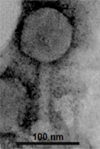 |
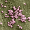
Ocean Bacteria Infected by Strange Looking Viruses
NCMIR researchers aided in the discovery of the most abundant virus in the ocean. This virus, called a Pelagiphage, attacks the most abundant microorganism in our oceans, a type of marine bacteria called SAR11, with significant implications for how carbon moves between the atmosphere and our oceans. SAR11, a bacterium that's the most abundant organism in the oceans, survives where most other cells would die and plays a major role in the planet's carbon cycle. It had been theorized that SAR11 was so small and widespread that it must be invulnerable to attack. Pelagiphages are so strange-looking that scientists previously didn't even recognize what they were. Read More > >
|
 |
Accumulated Excess Protein Linked To Development Of Parkinson’s Disease By Progressively Disrupting Neuronal Function And Viability
NCMIR scientists have shown that overexpression of a protein called alpha-synuclein appears to disrupt vital recycling processes in neurons, starting with the terminal extensions of neurons and working its way back to the cells’ center, with the potential consequence of progressive degeneration and eventual cell death. Published in the February 6, 2013 issue of The Journal of Neuroscience, this study has major implications for more fully understanding the causes and mechanisms of Parkinson’s disease (PD), a neurodegenerative movement disorder that affects an estimated 1 million Americans. Read More > >
|
 |
Electron Microscopy Achieves A New APEX
Chemists from MIT together with NCMIR Scientists, Mark Ellisman, Tom Deerinck and Gina Sosinsky, have now designed a GFP equivalent for electron microscopy — a tag that allows scientists to label and visualize proteins with unprecedented clarity that offers much higher resolution than fluorescence microscopy. The new tag developed by Jeff Martell and Alice Ting, lead and senior authors of a paper describing the new tag in the Oct. 21 online edition of Nature Biotechnology, could help scientists pinpoint the locations of many cell proteins, providing new insight into those proteins’ functions. This new tag can label proteins throughout the cell — not only within mitochondria but also in the nucleus, the endoplasmic reticulum and the cytosol. Read More > >
|
 |
It's Not What You Eat But When You Eat
Researchers at the Salk Institute led by Dr. Satchidananda Panda with NCMIR collaborators, published a provocative paper In the June issue of Cell Metabolism showing that mice fed a high calorie/high fat diet on a time restricted feeding schedule were protected against obesity and related complications compared to mice eating the same amount of calories ad libitum. Read More > >
|
 |
New Insights into the Role of LRRK2 Mutations in the Pathogenesis of Parkinson's Disease
Mutations in the leucine-rich repeat kinase 2 (LRRK2) are associated with increased risk of developing Parkinson’s disease. Kett and colleagues, in collaboration with researchers at NCMIR, have uncovered a novel association of mutant LRRK2 with microtubules using advanced labeling and microscopy techniques. In cultured neurons, LRRK2 mutations appear to enhanced LRRK2 oligomerization, causing it to form filamentous structures visible by light microscopy.
Read More > >
|
 |
Correlative Microscopy Webinar
n April 4, 2012, Mark Ellisman, NCMIR Director, participated in a webinar on “Correlative Microscopy” sponsored by the Biophysical Society, the Biophysical Journal and Cell Press. Moderated by Dr. Paul Wiseman, Associate Professor of Physics and Chemistry at McGill University, the topics in this webinar ranged from combining light, electron microscopy (both selectively stained thick section EM tomography or cryo-EM tomography) and soft X-ray tomography. Read More > >
|
 |
NCMIR Researchers Help Create a Powerful New Probe for Light and Electron Microscopy
In the April 5th issue of PLoS Biology, NCMIR scientists, in a collaboration with a team led by Nobel laureate Roger Tsien, introduced a new fluorescent probe for correlated light and electron microscopy that overcomes many of the limitations previously encountered by researchers seeking to image cells and tissues at high resolution. Read More > >
|
 |
UC San Diego's National Center for Microscopy and Imaging Research (NCMIR) Instrumental in Huntington's Disease Breakthrough
Imagine being able to prevent certain death of nerve cells in the brain and visualizing the feat with the latest in electron microscope technology. That's just what a team of biomedical researchers did, including UC San Diego's own National Center for Microscopy and Imaging Research (NCMIR), giving hope to Huntington's disease patients. Huntington’s disease is an inherited and incurable neurodegenerative disorder affecting 35,000 people annually and is caused by mutation of the gene encoding the huntingtin protein. Read More > >
|
 |
New Research Reveals Unexpected Biological Pathway In Glaucoma
In a study published in the Proceedings of the National Academy of Sciences, a team of researchers from the Kennedy Krieger Institute and four collaborating institutions, including the National Center for Microscopy and Imaging Research, identified a new and unexpected biological pathway that appears to contribute to the development of glaucoma and its resulting vision loss. Read More > >
|
 |
Serial Block-Face Scanning Electron Microscopy at the 2010 Microscopy and Microanalysis MeetingAt the 2010 Microscopy and Microanalysis meeting in Portland, Oregon, NCMIR scientists presented their work "Enhancing Serial Block-Face Scanning Electron Microscopy to Enable High Resolution 3-D Nanohistology of Cells and Tissues," a collaboration with researchers Roger Tsien's laboratory at UCSD. Read More > > |
 |
NCMIR Scientists Provide the First Glimpse of Synthetic Life
Pioneering geneticist J. Craig Venter and colleagues announced the creation of the first synthetic life form, Mycoplasma mycoides JCVI-syn1.0 at a press conference in Washington, D.C. and NCMIR researchers were entrusted with obtaining the first images of this new organism. Read More > >
|
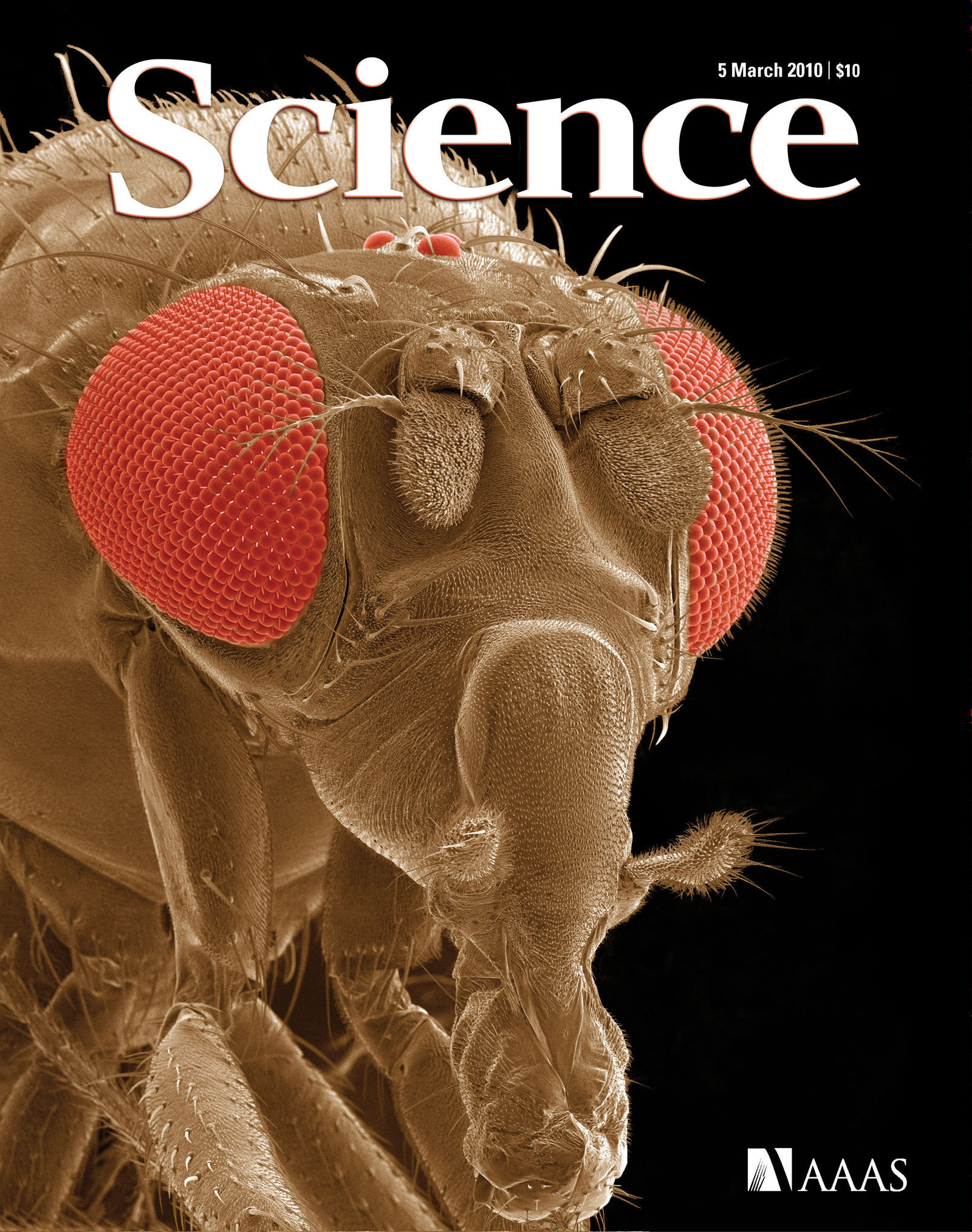 |
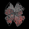
Sestrin Protects Fruit Flies From Age-Related Pathologies NCMIR Image Featured on Cover of Science
NCMIR scientists were members of a team of researchers that studied the intricate link of TOR with sestrins, a family of highly conserved proteins whose expression is induced by stress. Read More > >
|
 |
NCMIR’s Electron Tomography Resource Reveals Novel Mitochondrial Anchoring Scaffolds and Cristae Structured for High-rate Metabolism
Electron microscope tomography was used at the NCMIR to aid Prof. George Spirou (West Virginia University) in his investigation of the mitochondria-associated adherens complex (MAC) in the auditory brain stem and resulted in a publication in the J. Neuroscience. Read More > >
|
 |
Direct Restriction of Virus Release and Incorporation of the Interferon-Induced Protein BST-2 into HIV-1 Particles
In their paper, "Direct Restriction of Virus Release and Incorporation of the Interferon-Induced Protein BST-2 into HIV-1 Particles" published in PloS Pathogens, NMCIR researchers, in collaboration with Kathleen Fitzpatrick and John Guatelli of the UCSD School of Medicine show that the cellular protein BST-2 is involved in retaining newly formed HIV-1 virions on the surface of cells. Read More > >
|
 |
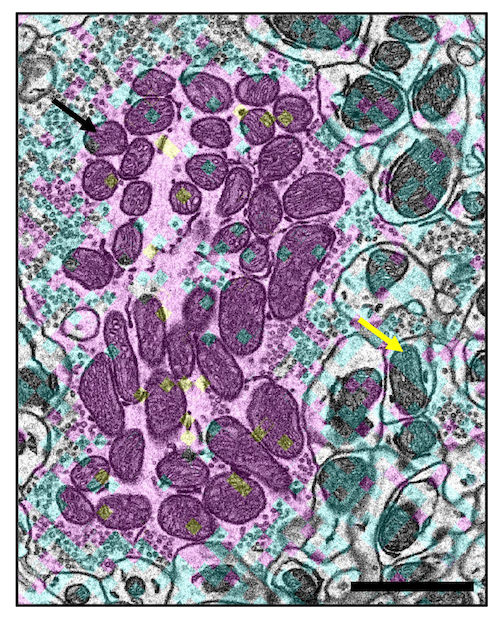
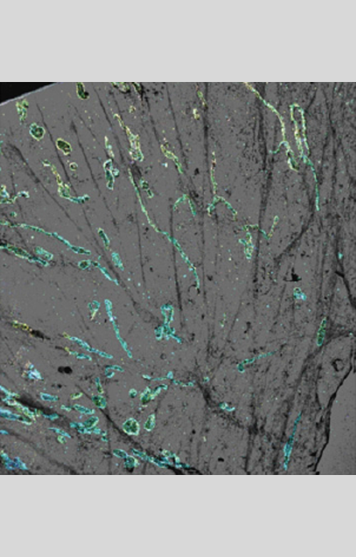
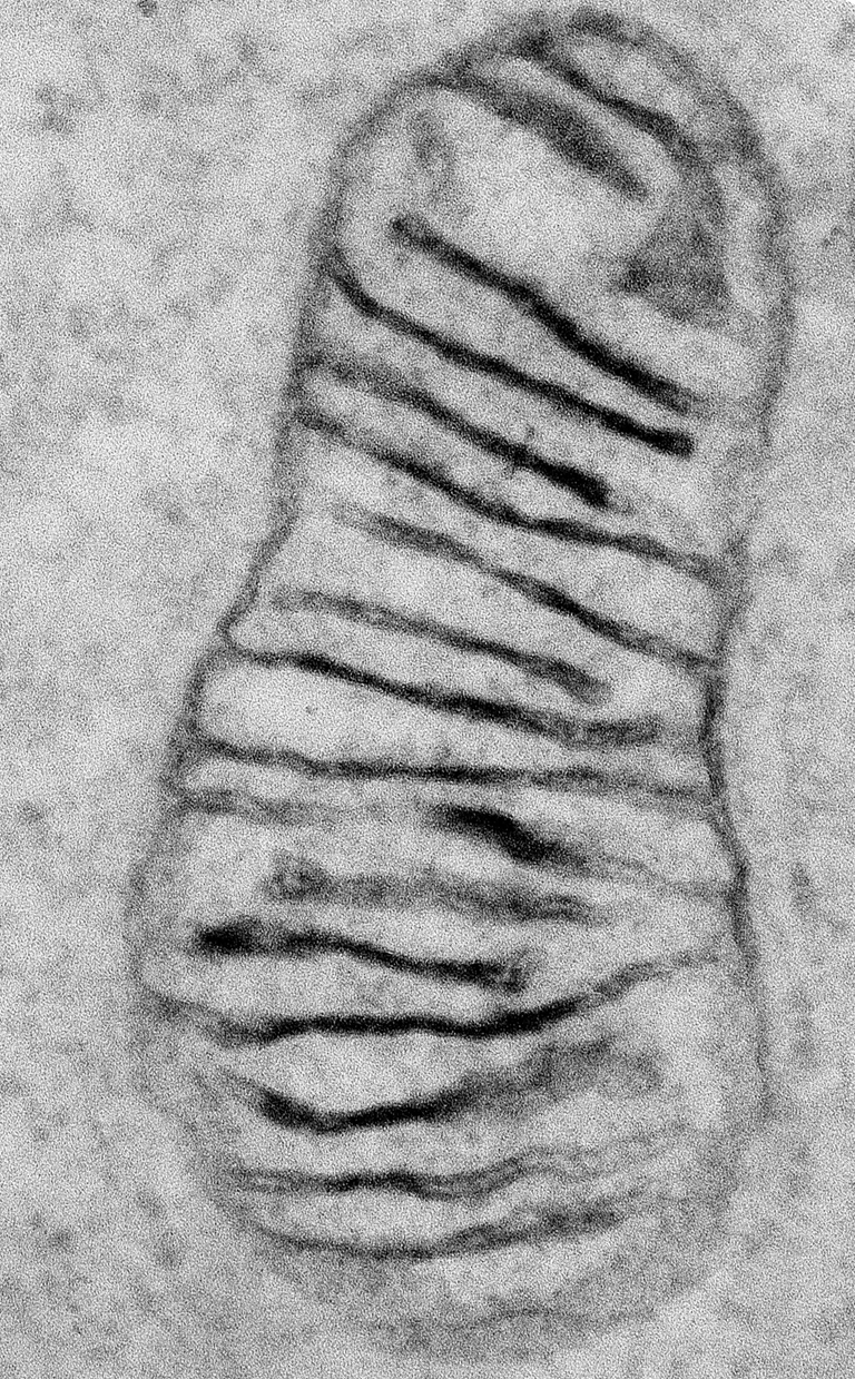
Mitochondrial configurations in peripheral nerve suggest differential ATP production.
NCMIR researchers used electron tomography to examine mitochondria, the cell’s principal energy producers, in the peripheral nervous system (PNS) in a study that points to differential energy usage along neurons. The morphological diversity of mitochondria in neurons appears to be the rule rather than the exception as these cells are highly polar, being composed of dendrites, somas, synapses, and axons — compartments with differing functions, molecules, and energetic needs. Read More > >
|
 |
New insights into mitochondrial structure during cell death
In this research effort, NCMIR neuroscientists and investigators at the Burnett School of Biomedical Sciences, College of Medicine, University of Central Florida, examined the pivotal role mitochondria play in the cascade of events associated with cell death pathways involved with several forms of neurodegeneration. Read More > >
|
 |
Three-dimensional electron microscopy reveals new details of membrane systems for Ca2+ signaling in the heart.
Using electron tomography, the NCMIR investigators in this study mapped the 3D topologies of dyadic clefts and associated membrane organelles in the mouse ventricular myocardium. Morphological details and the distribution of membrane systems, including transverse tubules (T-tubules), junctional sarcoplasmic reticulum (SR) and vicinal mitochondria, involved in controlling cardiac Ca2+ dynamics, were determined. Read More > >
|
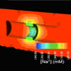 |
NCMIR scientists in combination with Salk Institute is the first to model axonal conduction to include a more realistic three-dimensional geometry of cellular microdomains.
Electrochemical signal transduction in cells depends on the subcellular organization of structural components, as well as on the electric profiles created by electrodiffusion of ions. Electrochemical diffusion is particularly important in nerve impulse transduction. With respect to neuronal cells and even at a higher systems level such as brain tissue, a fundamental question arises: how can seemingly similar cells such as neurons have such different electrophysiological properties from one another? Read More > >
|
 |
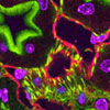
NCMIR collaborators demonstrate the use of fluorescent quantum dots for tracking dendritic cells and priming an immune response in vitro and in vivo.
Dendritic cells play a key role in initiating adaptive immune response by presenting antigen to T cells in lymphoid organs. NCMIR collaborators Michael Cahalan, Debasish Sen and Ian Parker at the University of California Irvine along with NCMIR scientists Thomas Deerinck and Mark Ellisman investigated the potential of using quantum dots as fluorescent nanoparticles for in vitro and in vivo imaging of dendritic cells, and as a particle-based antigen-delivery system to enhance denditic cell-mediated immune responses. Read More > >
|
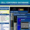 |
The CCDB Releases a New Version of Web Site to Make Sharing Microscopy Data Easier: http://ccdb.ucsd.edu
Originally deployed in March 2002 under the auspices of NIH’s Human Brain Project, the Cell Centered Database (CCDB) accommodates the storage and manipulation of high-resolution 3D light and electron microscopic reconstructions and facilitates advanced electron tomography.
Read More > > |
 |
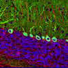
NCMIR collaborators reveal how new neurons, born in the hippocampus, wire into the brain’s circuitry
Just over a decade ago the idea that the mammalian brain contains stem cells that divide and differentiate into new neurons continually throughout life was largely unimaginable. However, with the recent body of research showing that areas of the adult brain continually undergo neurogenesis, neuroscientists have adopted a new paradigm in thinking about the brain. Read More > >
|
 |
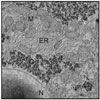
UCSD Researchers Reveal Dynamic Behavior of Golgi Complex
Researchers at UC San Diego reveal new details about the life cycle of the Golgi apparatus—the organelle responsible for correctly sorting, targeting, and trafficking protein in our cells. Constructing a novel protein tag suitable for light microscopy and correlated high-resolution electron microscopy, the team overcame intrinsic problems in observing the Golgi while undergoing cell division.
Read More > > |
 |
NCMIR researchers help biologists reveal greater structural detail.
In step with its mission of advancing the analysis of 3D structures in neurobiology and cell biology, a team of NCMIR researchers is helping biologists to take advantage of large-field electron microscope imaging developments for improving the resolution of 3D structures at the scale of proteins.
Read More > > |
 |
4th International Congress on Electron Tomography (4ICET)
NCMIR organized and hosted the Fourth International Congress on Electron Tomography (ICET) in San Diego on November 5-8, 2006. Convened three times since 1997, this conference provides a forum for structural and cell biologists, materials and computational scientists, and others to exchange information about how electron tomography (ET) elucidates structure-function in cellular arrays and larger assemblies. Read More > >
|
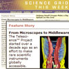 |
NCMIR's Telescience is featured in Science Grid This Week
The Telescience™ Project started over a decade ago as an effort to make rare scientific instruments globally accessible. Researchers at the National Center for Microscopy and Imaging Research began developing technologies to remotely control bio-imaging instruments over the Internet, and in 1992 the first system was demonstrated when attendees at a conference in Chicago interactively acquired and viewed images from one of NCMIR's intermediate voltage electron microscopes in San Diego. Read More > >
|
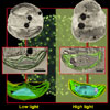 |
NCMIR’s electron tomography reveals ultrastructural details about how a phytoplankton adapts to changes in light intensity
Tiffany Moisan of NASA’s Goddard Space Flight Center at Wallops Island Virgina teamed up with NCMIR researchers to investigate the morphological changes associated with a marine alga, Phaeocystis antarctica, grown in light-limiting and high light environments. Their study, reported in the journal Marine Biology, presents the first 3D structures determined by electron tomography of an ecologically important phytoplankton species. Read More > >
|
 |
Studies of mouse models of human disease helped by NCMIR’s large-scale imaging expertise
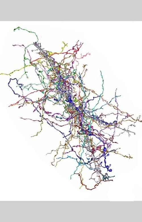
Scientists studying the nervous system have long struggled to understand how higher order structures, such as cellular networks, are assembled out of finer building blocks, such as dendritic and axonal architectures. Mapping the entire neural structure is the key to elucidating brain function. In a forthcoming issue of Neuroinformatics, a team of NCMIR researchers report on their progress towards this central goal by describing new procedures to study brain architecture and to characterize pathological changes in transgenic mice, spanning millimeter- to nanometer-sized structures. Read More > >
|
 |
NCMIR researcher wins second and eighth place in the Nikon International Small World Competition.
Nikon Corp has announced the winners of the 2005 Nikon International Small World Competition. Local NCMIR scientist Tom Deerinck garnered a second and an eighth place in the prestigious competition with his images of tissues stained with fluorescent quantum dots. According to Nikon: “The Nikon Small World competition recognizes the fusion of science and art as captured through the lens of the optical microscope. Read More > >
|
 |
Quantum Dots allow NCMIR scientists image cellular and subcellular detail from a single preparation.
Cell biologists at UCSD’s National Center for Microscopy and Imaging Research (NCMIR) demonstrate the suitability of Quantum dots (QDs) for determining the specific location of endogenous proteins in cells and tissues—using correlated light and electron imaging techniques. Read More > >
|
 |
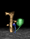
NCMIR and Salk Researchers “animate” synaptic transmission
In a collaboration between computational neuroscientists and neuroanatomists, researchers have created a 3-D, nano-scale “anatomically correct” neuronal synapse that provides new insights into the way synapses work. Efforts to model activities of the nervous system have generally required drastically simplifying the complex geometry of the brain and its microscopic components. Read More > >
|
 |
3D Mouse Model Reveals Complex Structures of the Node of Ranvier
NCMIR researchers, in collaboration with the Salk Institute, have created a 3D model of the node of Ranvier, a complex structure found in the nervous system of higher vertebrates. The results will be published in an upcoming issue of Neuroinformatics. Read More > >
|
 |
Tomography Method Improves Resolution in Thick Samples
A novel method for acquiring data for electron tomography dramatically increases resolution and usable section thickness of stained samples, according to a NCMIR study published in the December 2004 issue of the Journal of Structural Biology. Read More > >
|
 |
Astrocytes Limit CNS Regeneration
Investigators at Göteborg University and the University of Southern California found astrocytes play a significant role in limiting the regenerative capacity of the central nervous system. In the May 26, 2004 issue of the Journal of Neuroscience, researchers collaborating with NCMIR’s Eric Bushong demonstrated reactive astrocytes lacking intermediate filaments in mice show a limited hypertrophy of cell processes and synaptic regeneration in the hippocampus. Read More > >
|
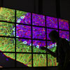 |
BioWall Displays Large Images of the Brain
NCMIR's newly installed "BioWall" tiled display, a 20-tile wall of high-resolution flat-panel displays that project massive, detailed 2D and 3D images of the brain, allows scientists to explore relationships among subcellular components, cells, and cellular networks at unprecedented resolution. Read More > >
|
 |
Nitric Oxide and Zinc Demolish Overexcited Neurons
A recent collaboration between NCMIR and the Center for Neuroscience and Aging at the Burnham Institute demonstrated a unique form of cross-talk between nitric oxide and zinc apoptotic signal transduction pathways that may contribute to neurodegeneration. Read More > >
|
 |
Purkinje Cell's Microenvironment May Be Regulated by Cerebellar Structure
A recent collaboration between NCMIR and the New York University Medical Center found that a cerebellar structure called the pinceau may regulate the microenviroment of the initial segment of the Purkinje cell axon. Read More > >
|
 |
Astrocytes Change During Postnatal Brain Development
Unique astrocyte territories in the adult brain are likely formed through competition between astrocyte processes, similar to the competitive interactions among neuronal dendritic fields, according to a recently published study by NCMIR researchers Eric A. Bushong, Maryann E. Martone, and Mark Ellisman that examined the fine morphological features of developing astrocytes in the rat brain. Read More > >
|
 |
NCMIR Scientist Places in Nikon's Small World Competition
NCMIR’s Tom Deerinck placed fourth in Nikon's 2003 Small World Competition with his image of a section of rat hippocampus stained with various cellular markers and imaged using 2-photon fluorescence microscopy. This is Deerinck's second win in this competition in as many years. Read More > >
|
 |
EM Tomography Emerges as a Useful Tool for Studying the Nervous System
These images, featured on the July 2003 cover of Microscopy Today, demonstrate the emerging importance of electron tomography as a research tool, particularly for studying the nervous system. Read More > >
|
 |
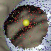
Relationship of Astrocytes to Laminar Boundaries in the Adult Brain Examined
The relationship of astrocytes to laminar boundaries in the adult brain was examined in a study published in the Journal of Comparitive Neurology. In this image, a protoplasmic astrocyte has been filled with a fluorescent dye (yellow) and the surrounding tissue is labeled for EphA4 (blue), revealing one of the laminar boundaries. Read More > >
|
 |
NCMIR Scientist Wins First in Nikon Small World Competition
Tom Deerinck’s photomicrograph of a sagittal section of rat cerebellum placed first in the 28th Annual Nikon International Small World Competition. Fluorescence and confocal microscopy were used to achieve this image. Read More > >
|
 |
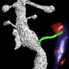
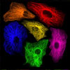
Connexin Trafficking Imaged Through Multicolor and Electron Microscopy
An optical section of double-labeled (FlAsH green and ReAsH red) gap junctions in HeLa cells is pictured. The tubulin network is shown in white, and the cell nuclei is shown in blue. Read More > >
|
 |
Synaptic Carbohydrate Antigen Plays Key Role in Neuromuscular Synapse Development
This laser scanning confocal image (BioRad Radiance confocal) is of a group of neuromuscular junctions from the mouse gastrocnemius muscle. The sample was triple labeled for acetylcholine receptors (red), synaptophysin (green), and neuron-specific enolase (blue). Read More > >
|
 |
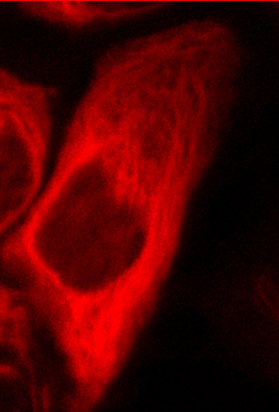
NCMIR Pioneers Use of Eosin for Fluorescence Photooxidation as a Staining Technique
A projection of a series of optical sections through a Purkinje neuron reveals both the overall morphology (red) and the dendritic spines (green). The unconjugated red dye fills the entire dendritic tree when injected, including shafts and spines. In contrast, the phalloidin-Alexa 488 conjugate selectively labels the spines. Read More > >
|
 |
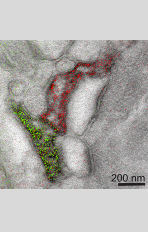
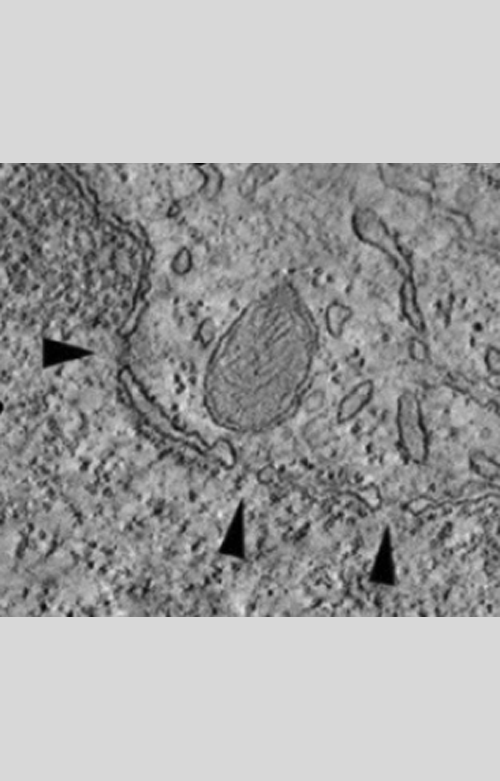
3D Image of Dendritic Spines Provides New Information on the Actin Cytoskeleton
Three-dimensional reconstruction of labeled spines using electron tomography showed that the labeled dense material was in continuity with the post-synaptic density. These results highlight differences in the actin cytoskeleton between different spine populations and provide novel information on the organization of the actin cytoskeleton in vivo. Read More > >
|