Research Highlights
Tomography Method Improves Resolution in Thick Samples
February 2005
A novel method for acquiring data for electron tomography dramatically increases resolution and usable section thickness of stained samples, according to a NCMIR study published in the December 2004 issue of the Journal of Structural Biology.
NCMIR investigator James Bouwer developed the automated method, known as “most-probable loss (MPL) tomography,” which takes advantage of a new generation of energy-filtering intermediate voltage electron microscopes (IVEMs), such as NCMIR’s JEM-3200EF IVEM (300 kV). MPL tracks a narrow bandwidth of electrons during tilt series acquisition. Instead of targeting zero loss electrons, as in most energy-filtering applications, MPL selects a narrow energy window where the maximum number of electrons are located.
As a section is tilted during tomography, the effective thickness that the electrons transverse increases. At higher tilts, almost all electrons undergo some loss of energy (inelastic scattering).
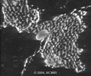
Bouwer, with co-authors Mason R. Mackey, Albert Lawrence, Tom Deerinck, Ying Jones, Masako Terada, Maryann E. Martone, Steven Peltier, and Mark H. Ellisman, also demonstrated that MPL increases signal-to-noise versus zero-loss imaging. As a result, MPL increases the usable thickness of selectively stained samples that can be imaged by IVEM to 3 um. This technique has also been extended to high voltage applications on the 1.25MeV microscope at the Korea Basic Science Institute, in South Korea. The figure of a rat hippocampus shows a maximum intensity projection through a tomographic volume of a 2 micron thick Golgi sample stained with a copper-lead stain and acquired at 1.25MeV.
For more details click here.