Research Highlights

Mitochondrial configurations in peripheral nerve suggest differential ATP production.

box
Condensed mitochondria in spinal root axons have a closely apposed outer and inner boundary membranes and narrow crista junctions. (a)–(d) Successive 4.4-nm thick slices through the tomographic volume of a spinal root axon prepared by HPF/FS. The matrix (m) is darker (more condensed) than the intracristal space (i). As is typical of condensed axonal mitochondria in peripheral nerve, there are few, albeit large, cristae. This example has only two cristae (1 and 2). Uncharacteristic of condensed mitochondria that have been isolated, yet typical of the in situ organelle, the outer and inner boundary membranes are in close apposition (between white arrows). Common for axonal mitochondria in the condensed configuration, the cristae are separated by a relatively narrow “bridge” of matrix (black arrows). Two crista junctions were observed in these slices, one per crista. Boxed areas were expanded 3x and placed as insets top and bottom. The top crista junction is barely open in (a), open in (b) and (c), and closed in (d). The bottom junction is closed in (a) and (d) and open in (b) and (c). The numbered, white-boxed areas in (c) are shown enlarged in (h) and are examples of contact sites between outer and inner boundary membranes. A portion of a microtubule next to the mitochondrion is outlined. scale bar = 200 nm. (e) and (f) Top view and side view of the segmented volume that was surface-rendered. The outer membrane is shown in blue and made transparent to see the two cristae (green and cyan). The visible crista junctions are indicated by arrowheads. (g) Segmented inner boundary membrane and side view of the surface-rendered volume. Seven crista junctions are seen in this view (numbered). The narrowness of the opening is similar to what is observed with orthodox mitochondria in situ, but less so with the isolated condensed organelle. See supplemental movie 1 for 3D perspectives. (h) Contact sites between the outer and inner boundary membranes are abundant. Boxed areas in (c) are shown enlarged 3x to demonstrate the range of membrane contacts (arrowheads). 1. A bridge contact spanning the intermembrane space. 2. Two bridge contacts close to each other with the top bridge being about twice as thick as the bottom bridge. 3. A classical contact site. 4. A classical contact site that is about twice as long. 5. An area where the outer and inner boundary membranes appear to be “zippered”. (i) Mitochondrial association with microtubules is greatest at the internode and least at the node. The percentage of mitochondria in close proximity to microtubules (e.g. see d), observed with both cTEM and electron tomography. The number of measurements is shown over each bar (*p < 0.05—applies to node/internode only). Six animals were used. Error bar = SEM for n = 6. (j) Crista junctions in condensed mitochondria are not enlarged. The mean crista width at its largest opening in tomographic reconstructions is compared in all axonal regions for orthodox and condensed mitochondria. The number of measurements is shown over each bar. Error bar = SEM.

December 2010

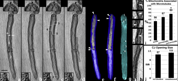
NCMIR researchers used electron tomography to examine mitochondria, the cell’s principal energy producers, in the peripheral nervous system (PNS) in a study that points to differential energy usage along neurons. The morphological diversity of mitochondria in neurons appears to be the rule rather than the exception as these cells are highly polar, being composed of dendrites, somas, synapses, and axons -- compartments with differing functions, molecules, and energetic needs. The abundance and conformational states of mitochondria in peripheral nerve were investigated because of energy considerations for action potential propagation in axons and especially the high metabolic load required to support ionic fluxes and phosphorylation and dephosphorylation of the molecular machinery (for opening and closing channels or binding to or unbinding from receptors or enzymes) concentrated at the node of Ranvier (a defined structure that governs how an action potential signal jumps along the axon, from node to node). Neuronal mitochondria display considerable structural diversity, particularly with respect to their cristae, the internal and unique membrane system governing ATP production, the primary molecule used as the energy currency of the cell. Evidence is accumulating that the topology of the cristae membranes is not random but rather is a regulated parameter. Cristae topology affects the diffusion of metabolites and soluble proteins that can influence mitochondrial ATP generation.  The condensed state of cristae corresponds to ATP generation several times higher than normal. An abundance of condensed mitochondria in PNS axons was reported with the density of mitochondria greatest at the node of Ranvier, supporting the concept that there is a need for the mitochondria to operate at a high workload of ATP production, especially at the node.  This type of mitochondrial state does not occur in the surrounding Schwann cells that generate the myelin wrappings around the axon or in central nervous system (CNS) axons, suggesting that the extreme length of PNS axons requires that mitochondria are transported and positioned such that proper functioning matches with maintaining high areas of ATP production in the most energy intensive axonal areas, i.e., the node of Ranvier. The positioning and energetic state of mitochondria appears to be essential for neuronal energy homeostasis.

Full text: http://www.ncbi.nlm.nih.gov/pubmed/20600951

Related Publications

Perkins GA, Ellisman MH. Mitochondrial configurations in peripheral nerve suggest differential ATP production. J Struct Biol. 2011 Jan;173(1):117-27. Epub 2010 Jun 25.