Research Highlights
Ultrastructure of Synaptic Connectivity within Subregions of the Suprachiasmatic Nucleus Revealed by a Genetically Encoded Tag and Serial Blockface Electron Microscopy
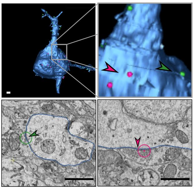
3D reconstruction of a neuron (upper panel) and EM images of synapses (lower panel) on a proximal dendrite (green arrow, left) and on soma (red arrow, right). Scale bar = 1 µm.
27-07-2023 La Jolla, CA
The hypothalamic suprachiasmatic nucleus (SCN) in the vertebrate brain serves as the central pacemaker regulating circadian rhythm throughout the body and as the principal hub for entraining circadian rhythm with the ambient light/dark cycle. The SCN receives photic information exclusively through melanopsin-expressing retinal ganglion cells (mRGCs) to synchronize circadian rhythms with the environmental light cycles. The SCN itself is composed of two major peptidergic neuron types in the core and shell regions of the SCN.
Cellular and molecular studies have suggested heterogeneity of the SCN neurons and their connectivity, yet an ultrastructural characterization of the SCN connectivity is still lacking. In order to systematically investigate the connectivity within the SCN, the investigators used a recently developed Cre-dependent electron microscopy (EM) reporter, APEX2, to specifically label mitochondria of melanopsin-expressing retinal ganglion cells (mRGCs), and serial blockface scanning EM (SBEM) to produce image volumes of the two functional subregions of the SCN, the core and the shell.
The resulting maps reveal patterns of connectomic organization in the core and shell of the SCN, in particular, showing that these regions are composed of different neuronal subtypes and differ with regard to the pattern of mRGC input, as the shell receives denser mRGC synaptic input compared with the core. This finding challenges the present view that photic information coming directly from the retina is received primarily by the core region of the SCN. Through this work, the team further established morphologic criteria for discriminating between different types of axonic boutons.
Original article at eNeuro:
Calligaro, Hugo, Azarin Shoghi, Xinyue Chen, Keun-Young Kim, Hsin Liu Yu, Brian Khov, Benjamin Finander, Hiep Le, Mark H. Ellisman, and Satchidananda Panda. "Ultrastructure of Synaptic Connectivity within Subregions of the Suprachiasmatic Nucleus Revealed by a Genetically Encoded Tag and Serial Blockface Electron Microscopy." Eneuro 10, no. 8 (2023).