Research Highlights
High-resolution volumetric imaging constrains compartmental models to explore synaptic integration and temporal processing by cochlear nucleus globular bushy cells
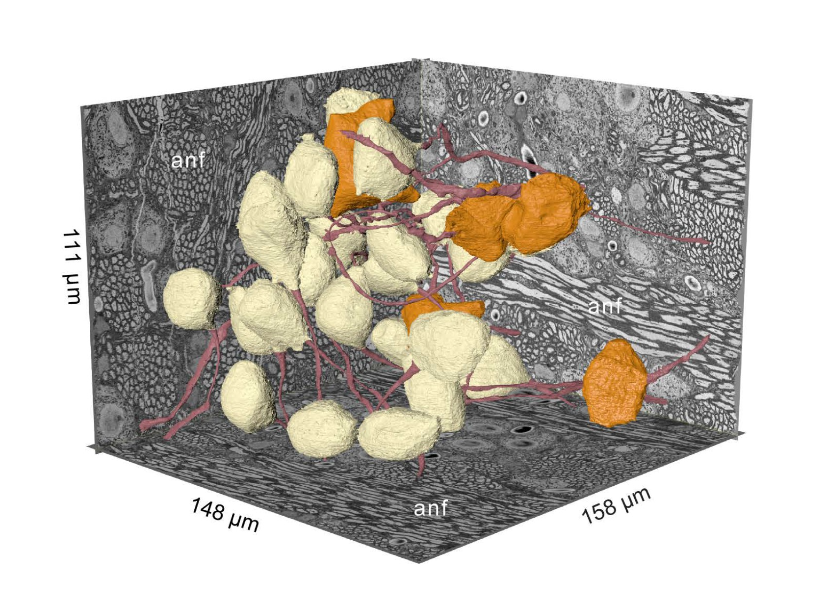
SBEM volume from the cochlear nucleus captures globular bushy (GBC) and multipolar cells (MC), and reveals synaptic sites. Twenty-six GBCs (beige) and 5 MCs (orange) are shown with their axons (red). Left rear wall transects auditory nerve (anf) fascicles, which run parallel to the right rear wall and floor. Non-anf axons exit into the right rear wall and floor as part of other fascicles that are cross-sectioned.
08-06-2023 La Jolla, CA
Globular bushy cells (GBCs) of the cochlear nucleus play central roles in the temporal processing of sound. Despite investigations spanning many decades, fundamental questions remain regarding their dendrite structure, afferent innervation, and integration of synaptic inputs.
Here, investigators use volume electron microscopy (EM) of the mouse cochlear nucleus to construct synaptic maps that precisely specify convergence ratios and synaptic weights for auditory nerve innervation and accurate surface areas of all postsynaptic compartments. Detailed biophysically based compartmental models can help develop hypotheses regarding how GBCs integrate inputs to yield their recorded responses to sound.
The team established a pipeline to yield precise reconstructions of auditory nerve axons and their endbulb terminals, together with high-resolution dendrite, soma, and axon reconstructions. These reconstructions create biophysically detailed compartmental models that could be activated by a standard cochlear transduction model.
With these constraints, the models predict auditory nerve input profiles where all endbulbs onto a GBC are subthreshold (coincidence detection mode), or one or two inputs are suprathreshold (mixed mode). The models also predict the relative importance of dendrite geometry, soma size, and axon initial segment length in setting action potential threshold and generating heterogeneity in sound-evoked responses, thereby proposing mechanisms by which GBCs may homeostatically adjust their excitability.
Volume EM also reveals new dendritic structures and dendrites that lack innervation. This framework defines a pathway from subcellular morphology to synaptic connectivity and facilitates investigation into the roles of specific cellular features in sound encoding. Through this work, the team also clarifies the need for new experimental measurements to provide missing cellular parameters and predict responses to sound for further in vivo studies, thereby serving as a template for the investigation of other neuron classes.
The original article in eLife:
Spirou, George A., Matthew G. Kersting, Sean Carr, Bayan Razzaq, Carolyna Yamamoto Alves Pinto, Mariah Dawson, Mark H. Ellisman, and Paul B. Manis. "High-resolution volumetric imaging constrains compartmental models to explore synaptic integration and temporal processing by cochlear nucleus globular bushy cells." Elife 12 (2023): e83393.