Research Highlights
Photoreceptor Disc Incisures Form as an Adaptive Mechanism Ensuring the Completion of Disc Enclosure
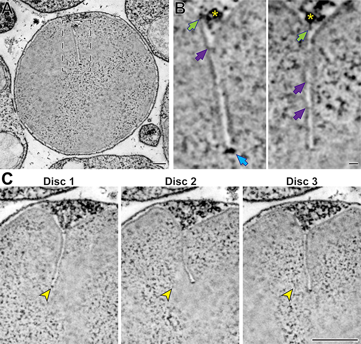
Ultrastructure of mature disc incisures. (A) Representative z-section from a reconstructed multi-tilt STEM tomogram of a 750-nm-thick WT mouse retinal section. (B) Representative maximum intensity projections of 3 (left) or 4 (right) z-sections highlighting various structures observed in a mature incisure. Green arrows point to structures spanning between microtubules and the disc rim; purple arrows point to connectors between opposing sides of the incisure; blue arrow points to the electron-dense structure at the incisure end. Yellow asterisk indicates the microtubule adjacent to the incisure. (C) Representative z-sections at the depths of 0 (Disc 1),+31.5 (Disc 2) and +63 nm (Disc 3) illustrating an imperfect alignment of incisures in three adjacent discs. Yellow arrowheads point to the incisure end in Disc 1 and the same x,y-coordinates in subsequent discs. Tomogram pixel size is 2.1 nm; scale bars: 0.25 µm (A, C) or 25 nm (B).
14-07-2023 La Jolla, CA
In the vertebrate retina, photoreceptor cells perform the first step of vision by capturing light and generating a neuronal signal. This process, called phototransduction, occurs within a stack of disc-shaped membranes, or 'discs,' confined within the ciliary outer segment. These discs contain deep indentations in their rims called 'incisures.' The presence of incisures has been documented in a variety of species, yet their role remains elusive.
In this study, the team combined traditional electron microscopy with three-dimensional electron tomography to demonstrate that incisures are formed only after discs become completely enclosed. They also observed that, at the earliest stage of their formation, discs are not round as typically depicted but are highly irregular in shape, resembling expanding lamellipodia.
Using genetically manipulated mice and frogs and measuring outer segment protein abundances by quantitative mass spectrometry, they further found that incisure size is determined by the molar ratio between peripherin-2, a disc rim protein critical for the process of disc enclosure, and rhodopsin, the major structural component of disc membranes. While a high peripherin-2 to rhodopsin ratio causes an increase in incisure size and structural complexity, a low ratio precludes incisure formation.
Based on these data, the investigators propose a model whereby normal rods express a modest excess of peripherin-2 over the amount required for complete disc enclosure to ensure that this important step of disc formation is accomplished. Once the disc is enclosed, the excess peripherin-2 incorporates into the rim to form an incisure.
The original article in eLife:
Tylor R Lewis, Sebastien Phan, Carson M Castillo, Keun-Young Kim, Kelsey Coppenrath, William Thomas, Ying Hao, Nikolai P Skiba, Marko E Horb, Mark H Ellisman, Vadim Y Arshavsky (2023) Photoreceptor disc incisures form as an adaptive mechanism ensuring the completion of disc enclosure eLife 12:e89160